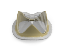
Jose L Navia and Sharif Al-Ruzzeh discuss the evolution of percutaneous technology and assess the technical limitations and challenges that need to be addressed in the percutaneous repair of aortic valves and mitral valves
“It is not the strongest of the species that survive, nor the most intelligent, but the one most responsive to change”, a famous quote by Charles Darwin that best describes how to deal with the present and future revolution that has been happening in the management of cardiac and vascular disease. There has been an accumulated development of percutaneous technology from the treatment of peripheral vascular disease by vascular surgeons and radiologists in the 50s and 60s, to percutaneous coronary intervention (PCI) for the management of coronary artery disease by cardiologists in the 70s and 80s, and to percutaneous management of thoracic and abdominal aortic disease by radiologists and cardiovascular surgeons in the 90s and 2000s.
All these differently trained physicians have been inadvertently helping evolve “intravascular percutaneous technology” in different domains and directions to treat vascular disease at different locations. This technology is now rising to a new horizon of treating intra-cardiac valves without conventional “open-heart” surgery.
Surgical valve repair (and/or replacement) is the gold standard treatment for valvular dysfunction and, therefore, newer approaches to managing valvular dysfunction must be at least as efficacious and have comparable safety data. However while open-heart procedures report short- and long-term successes, they are still associated with major morbidity and mortality, especially, when you consider the risks of reoperation for valve dysfunction, complications of thromboembolism and anticoagulation and endocarditis. All these factors have prompted clinicians to explore a variety of less invasive techniques including valve repairs, minimally invasive surgical approaches, and, more recently, percutaneous approaches toward valve repair or replacement.
Surgical treatment of mitral valve pathology has benefited from the evolution of surgical concepts. Refinement of repair techniques allows cardiac surgeons perform a successful repair in most patients with degenerative or ischaemic mitral insufficiency in experienced centres. Mitral valve repair may be more difficult in patients with rheumatic disease or infective endocarditis. Technological development has enabled surgeons to perform mitral valve surgery through limited incisions including ministernotomy, right minithoracotomy, and robotic mitral valve repair. All these approaches allow the surgeon to reproduce conventional surgical techniques, but through limited incisions that benefit patient recovery. For patients undergoing mitral valve replacement, preservation of the chordal apparatus preserves left ventricular function and enhances postoperative survival compared with mitral valve replacement with resection of the mitral apparatus.
The safety of this new technology demands certain basic features to be available including optimum imaging, such as echocardiography, new CT scan and MRI modalities, of the valve to be treated, hence the need of developed echocardiographic monitoring, the safe ablation of a special material valve and deployment of the valve within or without the native valve using a flexible delivery system; hence, the required development in biomedical sciences.
There are technical limitations and challenges of this evolving technology that are being addressed at present including, but not limited to, the suturing technique of the valve to the scaffold, the expandability of calcified tissue, and proper sizing in both the diameter of the valve and the length; the latter, perhaps more important for aortic valve procedures to avoid obstruction to coronary ostial flow. The preoperative assessment is crucial to guide selection and preparation of the stent-valve prosthesis. Industry has dealt with technical challenges like this in the past and undoubtedly it will succeed this time too.
Percutaneous and transapical implantation for aortic and pulmonary valved stents have shown promising results and are gaining acceptance in clinical practice. Several approaches have been explored to reproduce basic principles of mitral valve repair with transcatheter therapies. Positioning and securing a stented valve at the level of the atrioventricular valves posses several obstacles related to the anatomy of the mitral valve apparatus with chordae tendineae and papillary muscles that may interfere with complete expansion of the stent, and obstruction of the left ventricular outflow tract that may result from using the radial force of the stent inside the mitral valve annulus. Recent publications have reported initial success with transcatheter deployment of valved stents in the mitral position through a transapical approach, overcoming obstacles related to the subvalvular mitral apparatus. Additionally, several devices that can be delivered by transcatheter approach have been described to repair the mitral annulus or to optimise leaflet coaptation.
Jose L. Navia, staff cardiothoracic surgeon, and Sharif Al-Ruzzeh, staff project scientist,
Cleveland Clinic Foundation, USA. They are the editors of Percutaneous Valve Technology. Present and Future (Nova Publishers). For information, see: http://bit.ly/VSLfuq


