Fracturing the discs of dysfunctional mechanical aortic valves and placing a transcatheter valve in the vacant space offers new hope as an option for patients who previously had few options for treatment, according to new research.
Heart teams weighing up treatment options for patients with aortic valve disease know that not only must they seek to achieve the best result in the immediate term, but they must also plan for the long-term, with some patients living years, or potentially decades, into the future. Important considerations include how well the patient will tolerate invasive surgery; what the likelihood is that they will require future interventions; and how they will cope with the potential need for lifelong anticoagulation.
For patients aged 60 years or below, surgical aortic valve replacement (SAVR) with a mechanical valve has long been viewed as the gold-standard option for treating aortic stenosis due to the lasting durability of the valve. Studies point to mechanical valves having a lower risk of all-cause mortality than bioprosthetic valves in this younger cohort of patients, but their use comes with trade-offs. Patients face the need for lifelong anticoagulation after mechanical valve implantation, and if the valve fails—with reported reoperation rates of between 7–10% by 10 to 15 years—there are currently no minimally invasive options available, potentially necessitating further high-risk surgery. These issues have, over time, contributed to surgeons implanting more bioprosthetic valves—for which transcatheter valve-in-valve procedures are well established—with mechanical valve use shrinking.
However, new research suggests that a minimally invasive option for the replacement of failing mechanical valves may now be a reality, removing one of the major obstacles to more patients receiving mechanical valves in the first instance.
“If it [mechanical valve implantation] goes well, it’s the only surgery you have in your life; whereas with biological valves, you often have the need to have a second procedure if you are young enough,” interventional cardiologist Ignacio Amat-Santos (Hospital Clínico Universitario de Valladolid, Valladolid, Spain) tells Cardiovascular News, commenting that while the risk of needing a second intervention with a mechanical valve is uncommon, it is not an insignificant number of patients for whom treatment options are currently limited.
Amat-Santos and colleagues have dedicated much of their research over the last decade to finding a valve-in-valve replacement option for mechanical aortic valves, working on the hypothesis that mechanical valve dysfunction can be treated percutaneously via controlled disc dislodgement, followed by TAVI—a strategy known as valve-in-valve mechanical prosthesis (ViMech).
“We started by bench testing, putting mechanical valves in 3D models and trying to snare the discs to see what was easy to pull from them,” he explains. “That was not a good idea because it was very tough, and you block the disc for a long time—which haemodynamically is a bad thing for the patient.
“But then we realised that the discs made of pyrolytic carbon are like porcelain, which means they are strong, but they are fragile. Crossing in the middle of the two discs with a 12 or 14mm diameter balloon diameter peripheral balloon, we can very easily dislodge and fracture them into two or three fragments.”
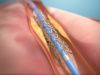
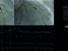
The technique they ultimately arrived at sees the discs of the valve fractured by the balloon, with the fragments then collected in an embolic filter to prevent them from passing through the blood stream and into critical organs. Once disc dislodgement has been confirmed on fluoroscopy, the filter and balloon are withdrawn, and a TAVI valve deployed immediately in the space vacated by the discs using the mechanical valve’s sewing ring as a landmark.
“It’s very easy to cross with the TAVI because we have created room, and the positioning of the TAVI is easy,” he comments. “We think it’s better to implant quite high to get good haemodynamics because the frame of the mechanical valves is quite rigid and often these are small prostheses.”
Taking their research from the bench to preclinical study, Amat-Santos and his team performed an animal study, implanting mechanical valves in pigs who underwent the ViMech procedure after one month. Via a bilateral femoral access route, the investigators implanted Ono Filters in the ascending aorta with a balloon positioned inside to fracture the discs, with another wire inserted for a TAVI device, using either Myval (Meril Life Sciences) or Evolut (Medtronic) valves.
The first tests in animals were encouraging, Amat-Santos tells Cardiovascular News, explaining that the researchers had planned to perform around 15 such procedures. However, after three, the team were presented with an emergent case in the hospital involving a patient with an embolised disc, which necessitated their first-in-man procedure. In a paper published in the European Heart Journal this January, the team details the first three human ViMech cases performed at their centre. The procedures, performed in patients aged 67–79 who had undergone multiple previous surgeries or had severe mechanical valve-related complications that made reoperation impossible, immediately restored valve function, with no major neurological or vascular events during follow-up, the team reports.
Amat-Santos says he hopes that publication of the report will stimulate further research in this area, and ultimately, convince more surgeons to consider implanting mechanical valves, with the knowledge that a ViMech procedure may be feasible down the line.
“Since we published this, several colleagues have been in contact with similar cases. In general, these are cases that can’t wait; one disc is not working and the patient is deteriorating slowly. I have done another case in Portugal, there is another case in Greece. And little by little, they are commenting on cases that potentially can be treated like this,” he says.
The research team has also created an online database and protocol to assist in collecting data on these procedures, as well as developing an app which has diameters of all mechanical valves as well as images of how they appear on fluoroscopy to aid in the implant of the TAVI valves.