
This content is only for readers outside of the USA
In an environment with growing demand for specialist cardiology services, it is important that health tech innovators support physicians to manage demanding workloads and complex procedures while ensuring the patient remains paramount. Within cardiac ultrasound solutions, recent developments are helping to drive improvements not only in terms of patient outcomes but also the user experience. When it comes to ensuring a confident diagnosis, for clinicians, every diagnostic image and patient moment in the care pathway matters.
So, where is the forward momentum heading?
Increasing the capacity for more
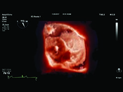
Fundamental step-change has been facilitated to improve performance overall via more powerful processing power of imaging hardware and software, offering advancing visualisation programmes—in some instances with photo-realistic quality. Users no longer have to choose between frame
rate and image quality. New technology more than doubles the frame rate without impacting image quality, offering focused images with fewer transmit operations to provide highly detailed ultrasound images and extraordinary temporal resolution. Automatic anatomical recognition, protocols for automatic functionality and proven quantification, make exams easier to perform and more reproducible, delivering new levels of clinical information.
This improved clinical information means faster and more consistent exams that are not only easier to perform but also provide clinicians with a high level of confidence, even for technically difficult patients, making it easier for cardiologists to quickly spot the warning signs of serious heart conditions and confidently assess when an interventional procedure is necessary.
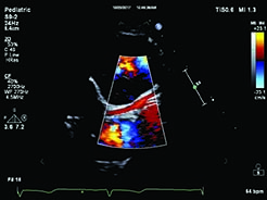
As technology evolves to capture better images across patient types and make the processes quicker and easier, it supports the treatment-planning processes through faster access to useful information. Recent developments and enhancements include transducers designed specifically for paediatric cardiology, tools which allow for multi-beat analysis from one acquisition, as well as advanced monitors and one-click sub-mode options. Developments like this support getting the best possible images for a confident diagnosis, even when visualising smaller vessels such as coronary arteries within paediatric patients.
Simplifying the user interface to advance the patient image
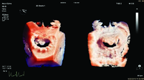
As well as image quality, the development of increasingly intuitive customisable user interfaces is a vitally important component of cardiology imaging and echocardiography. By simplifying controls, the clinician can focus on caring for the patient, while the ergonomics and portability, alongside truly configurable cardiac user interface and simplified echo guidance facilities, allows a streamlined workflow and optimised data capture consistency exam-to-exam, for robust decision making and ultimately resulting in improved exam efficiencies. It’s now possible to achieve better visualisation of cardiac structures for procedure guidance in fewer steps, confidently visualise the region of interest for echo-guided interventional procedures such as mitral valve repair, and obtain faster 3D measurements for device sizing.
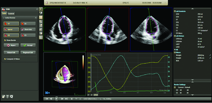
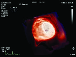
Jon Weait, Ultrasound Business Marketing Manager for UKI, Philips, said: “With the average age of the UK population on the increase, so is the prevalence of heart disease. At Philips, we are proud of the role we play in meeting these evolving population health needs and through meaningful innovation we continue to help create cardiology solutions that improve the patient and physician experiences. Central to this is the belief that richer images are key to ensuring accurate, efficient patient care in cardiology and in a recent simulated evaluation, with over 40 Philips users globally, 90%1 of clinicians felt the new TrueVue 3D photorealistic rendering improved viewing of anatomical structures, thus increasing clinical confidence, and 95%1 said they believe that the new EPIQ CVx cardiology ultrasound system with the latest OLED technology offers improved image quality in terms of sharper and clearer images.1 We are excited to be able to continue to improve the treatment landscape for cardiology professionals in order to provide the best patient care. Because every patient matters, every image counts”.
To find out more about Philips latest EPIQ CVx cardiovascular ultrasound system, click here
Reference
- These results were obtained during user demonstrations performed in December 2017 with the EPIQ CVx and the iE33 systems. The research was designed and supervised by Use-Lab GmbH, an independent and objective engineering consultancy and user interface design company. The tests involved 40+ clinicians from 17 countries. The various types of cardiac customer segments represented were adult diagnostics and interventional, adult diagnostics, and paediatric diagnostics and interventional.


