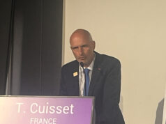
At the South Africa Heart Association annual congress (8–11 September, Cape Town, South Africa), Jacques Scherman (Chris Barnard Division of Cardiothoracic Surgery, Groote Schuur Hospital, University of Cape Town, Cape Town, South Africa) presented a proof-of-concept study for a novel, non-occlusive, self-locating transcatheter aortic valve implantation (TAVI) device. He talks to Cardiovascular News about why the device is a potential new option for patients with rheumatic heart disease.
Why is the incidence of rheumatic fever (ie. the cause of rheumatic heart disease) higher in developing countries than it is in developed countries?
Rheumatic fever and rheumatic heart disease are diseases of poverty and low socioeconomic circumstances—conditions that are often present in the developing world. Recent studies have found that in South Africa, the incidence of rheumatic heart disease is 24/100,000 per year among those aged more than 14 years. This is in stark contrast to the incidence in developed countries: <1/100,000.
More importantly, within 30 months of diagnosis, 22% of patients needed heart valve surgery.
It is estimated that in Africa alone there are 15 million patients living with rheumatic heart disease, of which 100,000 per year may need a heart valve intervention during some stage of their lives.
In developing countries, what percentage of patients requiring aortic valve replacement will have rheumatic heart disease?
In our own experience, during the last 10 years, rheumatic heart disease was the underlying pathology in more than 50% of patients requiring an isolated aortic valve replacement. Even in large urban centres in China and India, a significant proportion of heart valve patients have a rheumatic aetiology.
What are the current treatment options for patients with rheumatic heart disease (who require a new valve)?
Due to the nature of the underlying disease, aortic valve repair options are limited. As such, an aortic valve replacement remains the treatment option of choice for the majority of patients suffering from symptomatic rheumatic aortic valve disease.

What are the difficulties of performing TAVI in these patients?
In patients with degenerative calcific aortic valve disease, positioning and placing of the TAVI valve typically relies on the fluoroscopic “footprint” inherent to the underlying calcific process.
Rheumatic aortic valve pathology is usually non-calcific, therefore, making the implantation of a new TAVI valve using fluoroscopy and echocardiography unique and more challenging. Furthermore, conventional TAVI devices rely on the crushed hydroxyapatite deposits—seen in the typically older patients who undergo transcatheter procedures in the developed world—to “anchor” the valves. In patients with rheumatic heart disease, such calcium deposits are usually absent and, thus, alternative options for anchoring the device are needed when performing TAVI in these patients
How is the valve that you developed simplified compared with existing TAVI devices?
The currently available balloon expandable TAVI valves require the use of sophisticated imaging modalities and rapid ventricular pacing during valve implantation. With rapid ventricular pacing, a temporary pacemaker is used to let the heart beat so fast that there is no blood going to the rest of the body. This can only be tolerated for short periods and, therefore, limits or hastens the time available for implantation of the valve.
In a proof-of-concept study, performed as acute animal experiments, we successfully demonstrated the feasibility of a new non-occlusive, self-locating transapical TAVI delivery system, obviating the need for rapid ventricular pacing and simplifying transcatheter aortic valve replacement. Using tactile feedback, the device is stabilised in the correct position within the aortic root during the implantation. It also has a temporary backflow valve to prevent blood leaking backwards into the ventricle during the implantation of the new valve. All these factors together allowed for a slow, controlled implantation compared with the currently available balloon-expandable devices.
What further studies are planned?
A chronic animal study will commence in October 2016 evaluating valve performance and outcomes up to six-months following implantation. Ex-vivo, using a fatigue tester, this new valve have already completed 360 million cycles (FDA requirements are 200 million cycles, equating to five years in humans).
First-in-man studies are scheduled to commence in October 2017.
Could this valve potentially be used for patients with aortic stenosis?
The unique, self-locating and non-occlusive design allows for the potential use in most underlying aortic valve pathologies.


